Ratings & Reviews performance provides an overview of what users think of your app. Here are the key metrics to help you identify how your app is rated by users and how successful is your review management strategy.
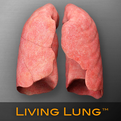
The Living Lung™ app is compatible with the iPad 2 or newer. Due to extremely high-resolution models and textures, this app is not compatible with the first generation iPads. OVERVIEW The Living Lung™ is a real-time 3D medical education and patient communication tool, featuring incredibly detailed anatomical respiratory models. It is a member of a series of apps developed specifically for the iPad by a team of anatomists, certified medical illustrators, animators, and programmers using actual human CT imaging data, and the most accurate 3D modeling technology available. The Living Lung™ is appropriate for use by secondary students, undergraduate and graduate students, and medical professionals. Interaction with the Living Lung™ utilizes true real-time 3D. Unlike some other anatomical apps and programs, there are no pre-rendered frames or animations. Therefore, the user can place the incredibly detailed lungs, rib cage, and associated structures in any position and zoom in to any location to explore the anatomical features. Animated transparent structures aid in the exploration of the bronchi and related key vascular components. Bronchopulmonary segments can be highlighted and labeled on the lungs and the bronchopulmonary tree. FEATURES Views: By selecting the views menu, the user can explore the anatomy of the lungs and associated structures by using a series of optional views. Color-coded, didactic models help to show the specific locations of lung segments, the internal bronchial tree, and circulatory structures. Lung Volume: By increasing or decreasing the breaths per minute, the user can observe the change in the lung volume. The results of the user interaction are shown in an accurate increase of decrease in the motion of the ribcage, inflation and deflation of the lungs and the associated internal structures. A lung volume meter shows the increased or decreased lung volume associated with the breaths per minute change. Labels: By selecting the color-coded segments of the bronchopulmonary tree, the user can study the names of the lung segments. The labels remain on screen and in the exact anatomical location during all real time 3D user interaction. About iSO-FORM iSO-FORM is a team of award winning medical artists, programmers and dreamers who believe that we are on the verge of a new era of learning, where the user doesn’t just memorize facts, but discovers them through interaction and curiosity. We love science, technology and art. That’s why iSO-FORM was born, so that we could live and work at that intersection. If we’ve piqued your curiosity, check us out at: www.iso-form.com.


